Spinal Fusion: Cervical
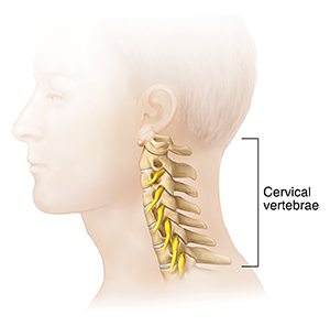
The cervical bones (vertebrae) are the top seven bones of your spine. They are located along your neck, just below the skull. Fusing vertebrae in the neck may help ease neck and arm pain. It may also help relieve progressive paralysis caused by compression of your nerve roots or spinal cord. Two or more vertebrae in your neck are fused. Fusion may be done through a cut in the front or the back of the neck. The surgery can take from 1 to 4 hours.

The fusion procedure

These steps apply to fusion from the front of the neck:
-
A head clamp or strap may be put on to align your neck.
-
The skin is cut and the muscles, blood vessels, trachea, and esophagus are pushed to one side to get to the vertebrae and disks.
-
An X-ray is done to check that the right spinal level is being operated on.
-
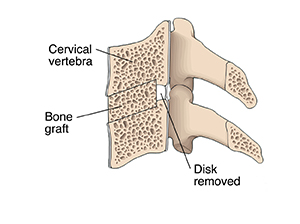
The disk is removed from between the vertebrae to be fused.
-
Bone spurs are removed.
-
A bone graft or intervertebral implant filled with bone is placed into the now-empty space between the vertebrae. Screws, plates, or a plate with screws are often placed in to ensure additional stability. In time, the graft and the bone around it will grow into a solid unit.
-
To help keep your spine steady and promote fusion, extra support with a cervical collar may be used.
-
A drain can be left in the wound for 1 or 2 days.
-
The incision is closed with stitches, glue, or staples.
-
A neck brace, rigid or soft, might be placed on your neck.
© 2000-2024 The StayWell Company, LLC. All rights reserved. This information is not intended as a substitute for professional medical care. Always follow your healthcare professional's instructions.